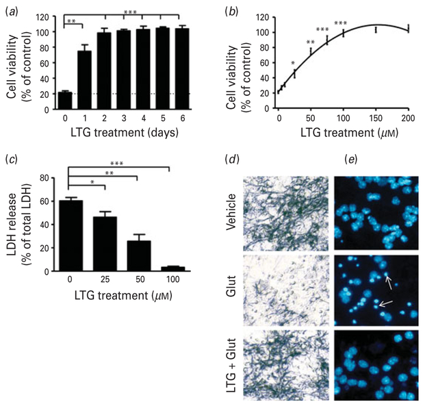
Fig. 1.
Pretreatment with lamotrigine (LTG) produced time and concentration-dependent protection against glutamate-induced excitotoxicity in cerebellar granule cells (CGCs). (a) Cells were pretreated with 100 μM LTG for the indicated number of days, prior to treatment with 100 μM glutamate (Glut) on 12 d in vitro (DIV) for 24 h. Cell viability was assessed by 3-(4,5-dimethylthiazol-2-yl)-2,5-diphenyl tetrazolium bromide (MTT) assay and expressed as % viability vs. vehicle-treated controls. (b) CGCs were treated with the indicated concentrations of LTG for 3 d, starting from 6 DIV, and then exposed to 100 μM Glut for 24 h. Cell viability was assessed by MTT assay and (c) lactate dehydrogenase (LDH) release expressed as %total LDH from lysed control cells. (d) Cells were stained with MTT for phase-contrast microscopy imaging or (e) Hoechst dye 33258 to detect chromatin condensation; arrows, apoptotic cells. Cell viability and LDH release are expressed as mean±S.E.M. from six independent cultures.* p<0.05, ** p<0.01, *** p<0.001 compared to Glut-only group.

