Figure 4.
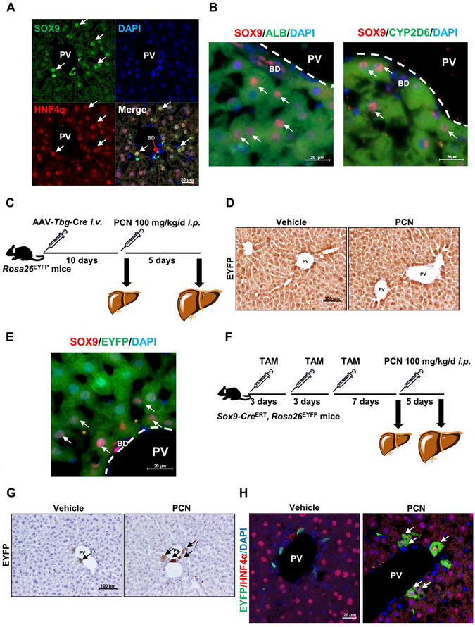
Clonal labelling confirmed cell type with PXR activation. (A) SOX9 and HNFα IF staining and DAPI staining in PCN-treated WT mice. (B) SOX9/ALB and SOX9/CYP2D6 double staining in PCN-treated mice. (C) Experimental design for TBG lineage tracing used Rosa26EYFPmice. Rosa26EYFP mice administered AAV-Tbg-Cre and treated with 100 mg/kg/d of PCN for 5 days. (D) Representative images showing EYFP staining. (E) SOX9 and EYFP double staining in PCN-treated AAV-Tbg-Cre, Rosa26EYFP ‘mice (n = 3). (F) Experimental design for SOX9 lineage tracing used Sox9-Cre, Rosa26EYFP mice administered TAM three times (once every three days), following treatment with 100 mg/kg/d of PCN for 5 days. (G) Representative slides of EYFP staining from Sox9-CreERT, Rosa26EYFPmice. (H) EYFP and HNF4a double staining in PCN-treated Sox9-CreERT, Rosa26EYFP mice (n = 3).

