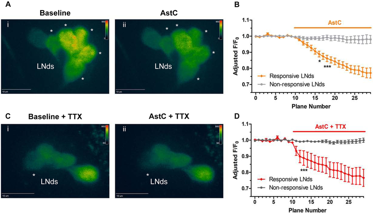
Figure 6. Functional calcium imaging of the LNds responding to AstC (10 μM).
Flies expressing the calcium sensor GCaMP6 in most clock neurons (clk856-GAL4> UAS-GCaMP6f) were collected in the evening, ZT9-12, and brains were explanted. A. A baseline recording of the LNds was obtained for 10 planes (Ai). When the synthetic AstC peptide (10 μM) was added into the bath, a substantial decrease in fluorescence was observed in multiple LNd neurons (Aii, denoted by asterisks). B. Quantification of the fluorescence relative to the initial baseline recording (F/F0) over time. Values were adjusted to remove trending bleach lines. The LNd neurons showed either a significant decrease in calcium (orange, ‘Responsive LNds’) or were unaffected (light gray, ‘Non-responsive LNds’, less than 10% change in fluorescence). C. To address whether AstC directly inhibited the LNds, the same experiment was conducted with the sodium channel blocker tetrodotoxin (TTX) added to the bath. Similar to (A), a baseline recording was obtained for the LNds (Ci) before exposing the brains to AstC (Cii). The asterisk denotes the single LNd neuron that showed a significant decrease in calcium in response to the AstC treatment. D. the quantification of the direct inhibition of AstC onto a single LNd neuron (red, ‘Responsive LNds’). Most LNd neurons were considered unaffected by having a less than 10% change in fluorescence. n = 6 brains per condition. * p < 0.05, *** p <0.001, two-way ANOVA.

