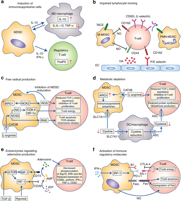
Fig. 3.
Main mechanisms of immunosuppression mediated by myeloid-derived suppressor cells (MDSCs). Mechanisms include the generation of immunosuppressive M2 macrophages and regulatory T cells via interleukin (IL)-10 and interferon (IFN)-γ secretion (a); impairment of lymphocyte adhesion to endothelial cells (ECs) and extravasation through nitric oxide (NO)-mediated downregulation of adhesion molecules CD162 and CD44, and tumor necrosis factor-alpha-converting enzyme (TACE)-mediated cleavage of CD62L (L-Selectin) on T cells (b); the production of reactive oxygen (ROS) and nitrogen species (RNS) through NADPH oxidase 2 (NOX-2) and nitric oxide synthase 2 (NOS2), leading to increased cyclooxygenase 2 (Cox-2), Hypoxia-inducible factor 1-alpha (HIF-1α) and arginase 1 (ARG1) expression and reduced T cell receptor (TCR) expression (c); the depletion and intracellular degradation of the amino acids L-arginine and cystine through increased uptake via the CAT2B and SLC7A11 transporters, respectively (d); induction of the ectoenzymes CD39 and CD73 via HIF-1 through transforming growth factor beta (TGF-β and hypoxic conditions, leading to adenosine production and reduced phosphorylation of extracellular signal–regulated kinase (ERK), protein kinase B (Akt) and Zap70, and reduced expression of CD95L, perforin, IFN-γ and tumour necrosis factor alpha TNF-α in T cells (e); and the expression of immune regulatory molecules B7, programmed death-ligand 1 (PD-L1) and FasL, causing T cell anergy and apoptosis via binding to their respective receptors (f)

