Fig. 5.
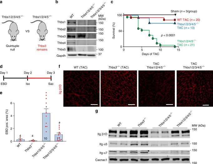
Induction of endogenous Thbs3 is detrimental after cardiac injury. a Schematic diagram depicting the Thbs gene-deleted mice used to selectively analyze effects of endogenous Thbs3. b Representative Western blots from hearts of 1–3 day-old WT, Thbs1/2/3/4/5−/− and Thbs1/2/4/5−/− mice for the indicated Thbs proteins. Gapdh served as loading control. c Kaplan–Meier survival plot of shams, WT, Thbs1/2/3/4/5−/− and Thbs1/2/4/5−/− mice after TAC surgery in days. Number of mice used is shown in the graph for each group. P < 0.0001 analyzed by log-rank test WT versus Thbs1/2/4/5−/− and Thbs1/2/3/4/5−/− versus Thbs1/2/4/5−/−. d Experimental regimen of EBD and Iso injection into the groups of mice shown (e) to measure membrane permeability. e Quantification of EBD positive area in the hearts of the indicated groups of mice after Iso injection with the regimen shown in d. *P < 0.05 versus WT; #P < 0.05 versus Thbs1/2/4/5−/− mice. Statistical analysis was performed using one-way ANOVA and Turkey multiple comparisons test. Error bars are +/− standard error of the mean and number of mice used in each experiment are shown in the graphs. f Immunohistochemistry for β1D integrin from heart sections of WT, Thbs3−/−, Thbs1/2/4/5−/− and Thbs1/2/3/4/5−/− mice 1 week after TAC surgery. Scale bars are 50 μm. g Representative Western blots for the indicated integrin proteins from hearts of WT, Thbs3−/−, Thbs1/2/4/5−/−, and Thbs1/2/3/4/5−/− mice subject to 1 week of TAC and processed for sarcolemma protein extracts. Cacna1c served as loading control. Quantitation of these results is shown in Supplementary Figure 4g–i

