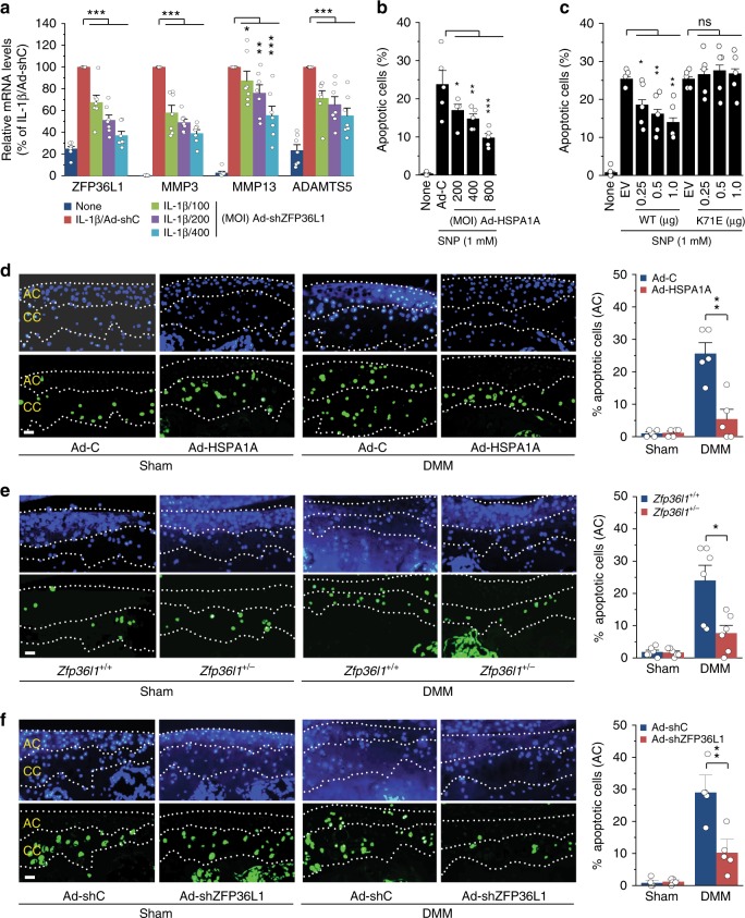
Fig. 6.
HSPA1A inhibits chondrocyte apoptosis. a qRT-PCR analysis (n = 7) of matrix-degrading enzymes in chondrocytes treated with IL-1β and infected with 400 MOI of control virus or the indicated MOIs of Ad-shZFP36L1. b, c Chondrocytes were infected with Ad-C or Ad-HSPA1A (b) or transfected with empty vector (EV, 1 μg) or vectors encoding WT-HSPA1A or K71E-HSPA1A (c) and left untreated or exposed to the NO donor, SNP, for 6 h. Apoptotic chondrocytes were identified and quantified by TUNEL staining (n = 5). d–f Detection and quantitation of apoptotic chondrocytes in calcified cartilage (CC) and articular cartilage (AC) of sham- or DMM-operated mice subjected to IA injection with Ad-C or Ad-HSPA1A (n = 5 mice per group) (d), sham- or DMM-operated Zfp36l1+/− and WT mice (e; n = 6 mice per group), or sham- or DMM-operated mice subjected to IA injection with Ad-shC or Ad-shZFP36L1 (f; n = 5 mice per group). Means ± s.e.m. with one-way ANOVA (a–c) and two-tailed t-test (d–f). *P < 0.05, **P < 0.005, and ***P < 0.0005; ns, not significant. Scale bar: 50 μm

