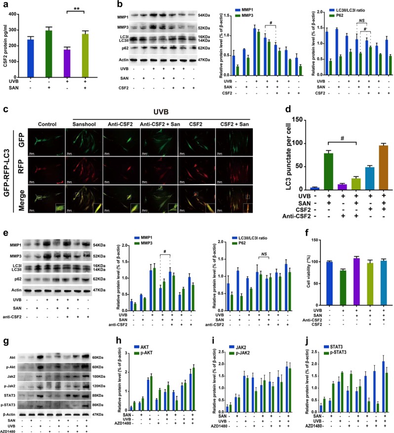
Fig. 3. Sanshool treatment induces autophagy and the CSF2-dependent JAK2–STAT3 signaling pathway in UVB-irradiated HDFs.
a HDFs treated with sanshool for 24 h after UVB irradiation; analysis of CSF2 secretion by ELISA; UVB-irradiated HDF or non-irradiated HDFs treated with sanshool when co-cultured with CSF2 (0.1 ng/mL). b Expression levels of LC3I/II, p62, MMP-1, MMP-3, and β-actin measured by Western blot analysis. c Representative images of LC3 staining by measuring fluorescence intensity in HDFs in different groups of cells infected with the RFP-GFP-LC3 adenovirus for 24 h by fluorescence microscopy. d Quantitation of punctate structures per cell, based on the number of punctate structures in GFP-RFP-LC3-positive cells. Addition of a CSF2-neutralizing antibody (5 μg/mL) to the medium in each group and incubation with sanshool in UVB-irradiated HDFs; e Expression levels of LC3I/II, p62, MMP-1, MMP-3, and actin measured by Western blot analysis; f Cell viability in response to CSF2 depletion or CSF2 oversupply. g Expression levels of JAK2, phospho-JAK2, STAT3, phospho-STAT3, AKT, and phospho-AKT after sanshool and/or AZD1480 treatment of UVB-irradiated or non-irradiated HDFs for 24 h, determined by Western blot analysis. (h) Relative protein expression levels of AKT and phospho-AKT. i Relative protein expression levels of JAK2 and phospho-JAK2; j Relative protein expression levels of STAT3 and phospho-STAT3. Results presented as means ± SD of three independent experiments (n = 3). *Compared with the UVB-treated group, P < 0.05

