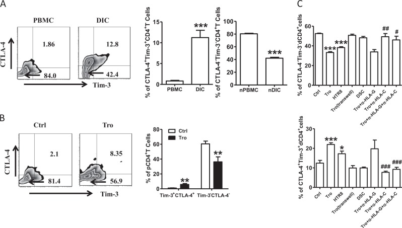
Fig. 3. Expression of CTLA-4 and Tim-3 on CD4+T cells during human early pregnancy.
a Flow cytometric analysis (left) and quantification (right) of CTLA-4 and Tim-3 co-expression on gated CD4+T cells from peripheral blood mononuclear cells (PBMCs) and decidual immune cells (DICs) during human normal first trimester pregnancies (n = 26).***P < 0.001. The flow cytometry plots were representative of three independent experiments. b Quantification of flow cytometric analysis of CTLA-4 and Tim-3 co-expression on peripheral CD4+T (pCD4+T)cells with or without co-culture with trophoblasts (Tros) for 48 h. n = 13. ***P < 0.001. d Quantification of flow cytometric analysis of CTLA-4 and Tim-3 expression on dCD4+T cells cultured alone or co-cultured with equal numbers of Tros (directly or indirectly), or human HTR8/SVneo cells, or decidual stromal cells (DSCs). The α-HLA-G and/or α-HLA-C antibody were used in some wells. n = 11, *P < 0.05, **P < 0.01, ***P < 0.001, Compared with the control. #P < 0.5, ##P < 0.01, ###P < 0.001, compared with the group co-cultured with Tros. Data represented the mean ± SEM

