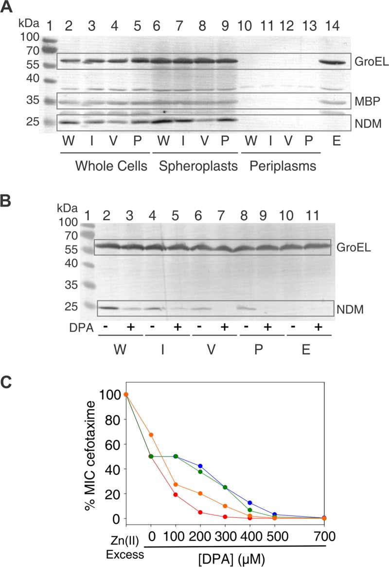
FIG 2.
Expression levels and Zn(II) limitation susceptibility of NDM-1 and L3 variants. (A) Immunoblot demonstrating steady-state expression in E. coli DH5α. Proteins were detected from whole-cell lysates (lanes 2 to 5 and lane 14), spheroplasts (lanes 6 to 9), and periplasmic extracts (lanes 10 to 13). Wild-type NDM-1 (W) corresponds to lanes 2, 6, and 10; L3IMP (I) corresponds to lanes 3, 7, and 11; L3VIM (V) corresponds to lanes 4, 8, and 12; L3Pro (P) corresponds to lanes 5, 9, and 13. Empty plasmid (E) corresponds to lane 14. Lane 1 shows the protein ladder marker. The GroEL molecular weight is 60 kDa and that for MBP is 47 kDa. (B) Immunoblot demonstrating steady-state expression of wild-type NDM-1 and the L3 variants in E. coli DH5α treated with DPA. After induction, cells were incubated with (+) or without (–) DPA, and protein expression was detected from of whole-cell lysates. Wild-type NDM-1 (W) corresponds to lanes 2 and 3, L3IMP (I) corresponds to lanes 4 and 5, L3VIM (V) corresponds to lanes 6 and 7, L3Pro (P) corresponds to lanes 8 and 9, and empty plasmid (E) corresponds to lanes 10 and 11. Untreated cells were loaded before treated ones. Lane 1 shows the protein ladder marker. (C) Antimicrobial susceptibility profiles of E. coli DH5α/pMBLe producing β-lactamases against cefotaxime at increasing DPA concentrations. E. coli DH5α expressing NDM-1 is shown in blue, E. coli DH5α expressing L3IMP is shown in green, E. coli DH5α expressing L3VIM is shown in red, and E. coli DH5α expressing L3Pro is shown in orange.

