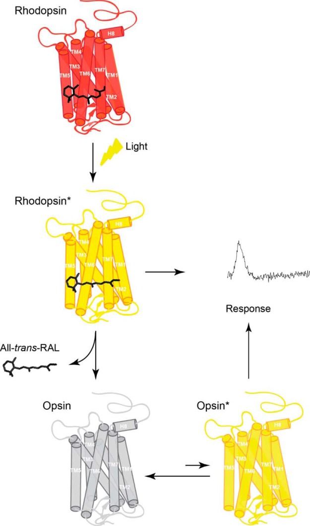
Figure 7.
Model of the mechanism of activation of rod phototransduction by free opsin. The chromophore 11-cis-retinal binds to apoprotein opsin via a protonated Schiff base in the retinal binding pocket forming the visual pigment (rhodopsin). Exposure of rhodopsin to light illumination results in the isomerization of 11-cis-retinal to all-trans-retinal, followed by conformational changes within protein, leading to formation of the receptor photoactivated state (rhodopsin*). Eventually, all-trans-retinal chromophore dissociates from the binding pocket of the short-lived rhodopsin*, resulting in the formation of unliganded opsin and free all-trans-retinal. Such ligand-free opsin exists in equilibrium between a predominant inactive opsin (opsin) and rare active opsin (opsin*). Both photoactivated rhodopsin* and opsin* generate a cellular response that can be recorded.

