
Figure 8.
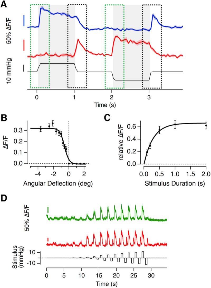
Hair cells signaling a return to rest. A, Example of iGluSnFR responses of two hair cells (not from the same recording), that generate a rebound-response; a large and transient release of glutamate after the release of a deflection in the null direction. B, The relationship between angular deflection in the null direction and amplitude of the rebound response. Data were averaged from 33 hair cells in which the largest rebound response exceeded 20% of the maximum response in the preferred direction (Rmax = 0.32 ± 0.01, Rmin = 0 ± 0.01, X1/2 = −0.56 ± 0.05 and Xs = 0.3 ± 0.05). C, The relationship between duration of deflection in the null direction and the magnitude of the rebound response (as a fraction of the response to stimulation in the preferred direction). These experiments were performed with large deflections generating the maximum rebound. Results are described by an exponential that yields a time constant of 0.3 s for the development of the rebound response. D, Two hair cells within the same neuromast, one of which (red) displaying a strong rebound response, whereas the other (green) does not, despite experiencing the exact same cupula deflection The relationship between cupula deflection and applied pressure in this neuromast was 0.29 deg/mmHg.