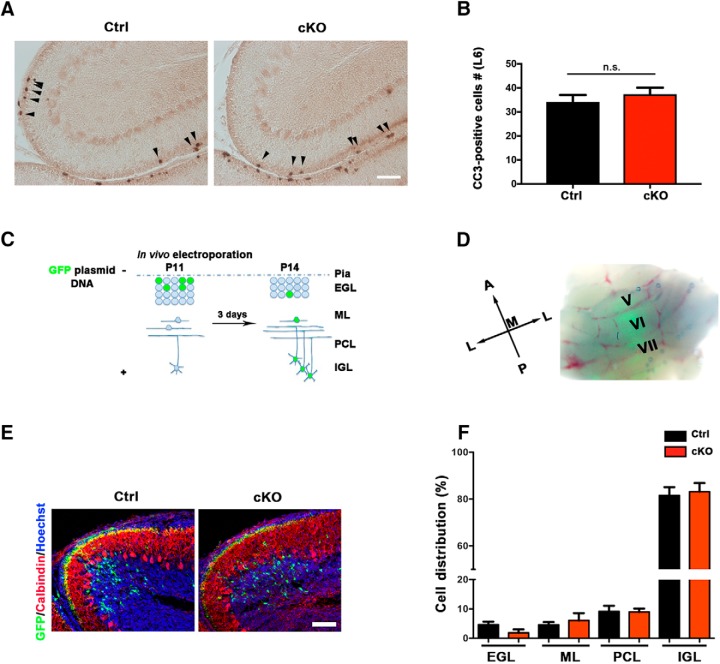
Figure 3.
Conditional SnoN KO in granule neuron precursors has little or no effect on apoptosis or migration of granule cells. A, The cerebellum from P14 conditional SnoN KO and control littermate mice was subjected to immunohistochemical analyses using the cleaved caspase-3 analyses. Scale bar, 100 μm. B, Quantification of the number of cleaved caspase-3-positive cells within the EGL in lobule VI revealed that conditional KO of SnoN had little or no effect on cell death in the EGL. Graphical data are presented as mean ± SEM, and significance determined by t test (n = 5 mice). C, A schematic representation of in vivo electroporation. D, The cerebellum of P11 mice electroporated with a GFP expression plasmid, which targets granule cell precursors, was analyzed at P14 by fluorescence microscopy. Robust GFP fluorescence was evident. E, The cerebellum of conditional SnoN KO and control littermate mice was subjected to immunohistochemical analyses using an antibody to GFP. GFP-labeled granule neurons were similarly positioned in conditional SnoN KO and control littermate mice. Scale bar, 100 μm. F, Quantification of the percentage of GFP-labeled granule cells within the EGL, molecular layer (ML), Purkinje cell layer (PCL), and IGL revealed that conditional SnoN KO had little or no effect on the positioning of granule neurons. Graphical data are presented as mean ± SEM, and significance determined by ANOVA followed by Fisher's PLSD post hoc test (n = 7 mice).

