Figure 4.
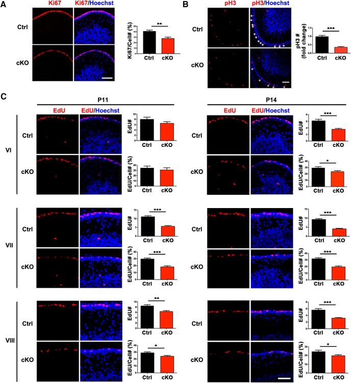
SnoN promotes proliferation of granule neuron precursors in the postnatal cerebellum. A, The cerebellum from P14 conditional SnoN KO and control littermate mice was subjected to immunohistochemical analyses using the Ki67 antibody and the DNA dye bisbenzimide (Hoechst). Substantially fewer Ki67-positive granule cell precursors were found in the EGL of conditional SnoN KO mice compared with control littermate animals. Scale bar, 50 μm. Graphical data are presented as mean ± SEM. **p < 0.01 (t test). n = 4 mice. B, The cerebellum from P14 conditional SnoN KO and control littermate mice was subjected to immunohistochemical analyses using an antibody to the pH3, which labels cells specifically during mitosis. Substantially fewer pH3-positive granule cell precursors were found in the EGL of conditional SnoN KO mice compared with control littermate animals. Scale bar, 50 μm. Graphical data are presented as mean ± SEM. ***p < 0.001 (t test). n = 4 mice. C, P11 and P14 conditional SnoN KO and littermate mice were injected intraperitoneally with the nucleotide derivative EdU. Thirty minutes after EdU injection, the cerebellum was subjected to histochemical analyses using an EdU detection reagent and the DNA dye bisbenzimide (Hoechst). The total number of EdU-labeled proliferating granule precursors and their proliferation index (percentage of EdU-labeled cells among all EGL cells) was significantly reduced in lobules VI, VII, and VIII at P14 and VII, VIII at P11 upon conditional KO of SnoN. Scale bar, 50 μm. The measured area equals 1000 μm2. All analyses were performed on three midline sections per brain. Graphical data are presented as mean ± SEM. ***p < 0.001 (t test). **p < 0.01 (t test). *p < 0.05 (t test). n = 4 mice.

