Figure 6.
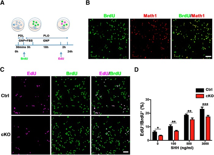
SnoN promotes proliferation of granule neuron precursors cell-autonomously and independently of SHH. A, A schematic of preparation of primary granule neuron precursors from P6 cerebellum. BrdU was used to label proliferating granule neuron precursors immediately after isolation, and 22 h later, a 2 h pulse of EdU incorporation was used to trace granule neuron precursors that remained in the cell cycle or those that exited the cell cycle. PDL, Poly-d-lysine; PLO, poly-ornithine; GNP, granule neuron precursor culture medium. B, BrdU-labeled proliferating granule neuron precursors were subjected to Math1 immunofluorescence. BrdU-positive cells almost completely colabeled with Math1, indicating the identity of mitotic granule neuron precursors. Scale bar, 50 μm. C, BrdU-labeled proliferating granule neuron precursors were subjected to EdU counterstaining. The percentage of primary granule neuron precursors that remained in the cell cycle (EdU+/BrdU+) was reduced upon conditional SnoN KO. Scale bar, 50 μm. D, Quantification of the percentage of EdU+/BrdU+ cells in control and conditional SnoN KO primary granule neuron precursors showed SHH dosage-dependent expansion. Exposure to SHH robustly increased proliferation of both control and SnoN-deficient granule neuron precursors, and SnoN depletion significantly reduced the rate of proliferation of granule neuron precursors in the presence or absence of SHH. Graphical data are presented as mean ± SEM. ***p < 0.001 (ANOVA followed by Fisher's PLSD post hoc test). **p < 0.01 (ANOVA followed by Fisher's PLSD post hoc test). *p < 0.05 (ANOVA followed by Fisher's PLSD post hoc test). n = 3 biological replicates.

