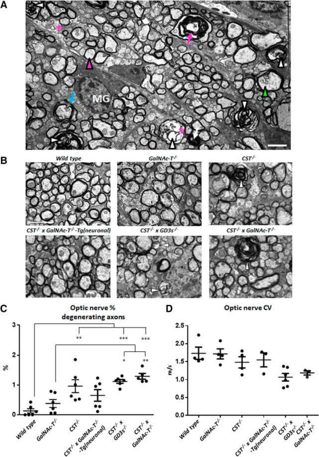
Figure 6.
Glycolipid deficiency causes pathological changes and compromises CNS axon survival and function. A, Representative ultrastructural features observed in CST−/− × GD3s−/− and CST−/− × GalNAc-T−/− mice. Shown are normal myelinated axon (magenta arrowhead), degenerating axons (white arrowheads), dark condensed degenerating axon (white arrow), vacuoles within axons (*), empty myelin sheath (magenta arrow), redundant myelin (blue arrow), abnormal cytoskeleton (green arrowhead), and microglia (MG). B, Electron micrographs depicting the differences among the genotypes. White arrowheads indicate degenerating axons. Myelin is similar among mouse lines. C, Degenerating axon number increases to a significant level compared with WT in OpNs from CST−/−, CST−/− × GD3s−/−, and CST−/− × GalNAc-T−/− mice. Reintroducing a- and b-series gangliosides into neurons rescues this pathology. D, Reduction in conduction velocity was observed in OpNs with diminishing glycolipid content. This did not reach significance between genotypes. One-way ANOVA followed by Tukey's post hoc tests to compare multiple comparisons, indicated on the graphs as follows: *p < 0.05; **p < 0.01; ***p < 0.001. Scale bar, 2 μm.

