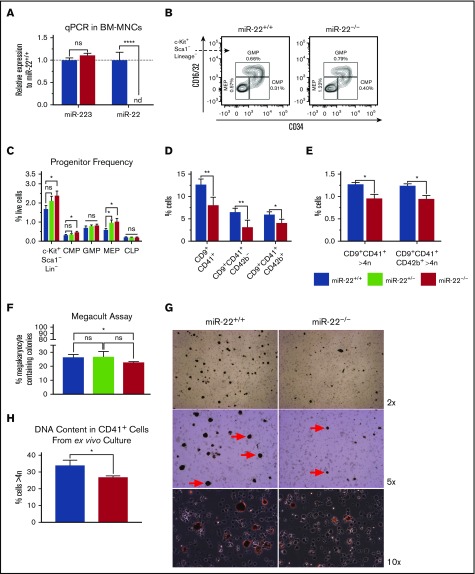
Figure 4.
miR-22 knockout mice exhibit an expansion of MK-erythrocyte progenitors and a defect in MK maturation. (A-C) Bone marrow from adult 129SV;miR-22 wildtype, heterozygous, and homozygous knockouts was isolated and subjected to analysis for gene expression, flow cytometric analysis, and ex vivo megakaryocytic differentiation. (A) Total RNA was isolated from bone marrow from wildtype and miR-22KO animals. RT was carried out using miRNA-specific primers. miR-223 was assayed as a control. sno202 was used as a housekeeping gene to quantify relative expression between samples (n = 3). (B) Bone marrow mononuclear cells from individual mice were stained for progenitors (CLP, CMP, GMP, and MEP) according to surface markers listed in supplemental Table 3, and DAPI for live/dead assessment. Shown are representative flow cytometry plots gated for myeloid progenitors from c-Kit+Sca1−Lineage− cells in wildtype and miR-22KO animals. (C) Quantitation of flow cytometric analysis of hematopoietic progenitor cells from wildtype, heterozygous, and miR-22KO animals (n = 6-9). (D-E) Bone marrow mononuclear cells from individual mice were stained for immature (CD9+CD41+CD42b−) and mature (CD9+CD41+CD42b+) MKs, and for DNA content. Quantitation of frequency of immature and mature MKs (D), and quantitation of the frequency of high-ploidy cells in mature MKs (n = 3) (E). (F) CFU-MK assays. One thousand KSL were isolated from individual wildtype, heterozygous, and miR-22KO animals by FACS and were plated in 1.7 mL MegaCult supplemented with collagen and cytokines and plated in covered chamber slides for culture. After 7 days, cultures were dehydrated in acetone and stained for acetylcholinesterase and counterstained with Harris’ hematoxylin. CFU-MK and non-MK were quantified by a blinded counter on 2 separate days, and counts were averaged (n = 3). (G-H) Ex vivo MK differentiation of primary miR-22KO bone marrow cells. Bone marrow mononuclear cells were isolated from individual adult 129SV:miR-22 wildtype and miR-22KO and were subjected to ex vivo MK differentiation by treatment with TPO. Whole cultures were used for flow cytometry and acetylcholinesterase staining. (G) Representative microscopic images of acetylcholinesterase-stained cytospins from unfractionated ex vivo MK differentiation cultures. MKs are stained brown. Red arrows show MKs at 5× magnification. (H) Frequency of CD41+ cells with high DNA content (>4 n) in ex vivo differentiated MKs (n = 2-3). CFU-MK, colony forming unit–megakaryocyte; CLP, common lymphoid progenitor; CMP, common myeloid progenitor; GMP, granulocyte-monocyte progenitor; MEP, megakaryocyte-erythrocyte progenitor; nd, not detectable. *P ≤ .05; **P < .01; ****P < .0001.

