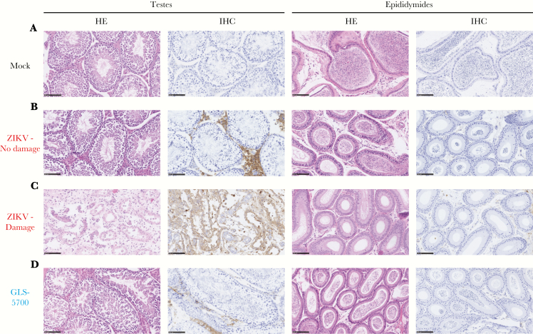
Figure 4.
Histopathological and immunohistochemical analysis of the testes and epididymides in male IFNAR−/− mice. (a) Histology and immunohistochemistry (IHC) of a representative testicle and epididymis of mock IFNAR−/− mice (mouse 1–7). (b) Histology and IHC of a representative testicle and epididymis of Zika virus (ZIKV)-infected IFNAR−/− mice that did not experience any damage to reproductive organs (mouse 2–13). (c) Histology and IHC of a representative testicle and epididymis of ZIKV-infected IFNAR−/− mice with damage to reproductive organs (mouse 2–11). (d) Histology and IHC of a representative testicle and epididymis of GLS-5700-vaccinated IFNAR−/− mice (mouse 3–7). All photographs were obtained from the left testis of each animal collected on day 28 postinfection and taken at a magnification of ×200; Scale bar = 100 µm. Abbreviation: HE, hematoxylin and eosin staining.

