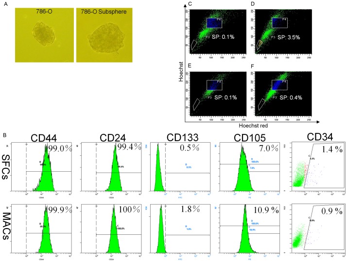
Figure 1.
Tumor spheres derived from 786-O cells share relevant features with stem cells. (A) Examples of primary spheres in 786-O and subspheres re-formed after single 786-O sphere cells were re-seeded at a clonal density and grown in DMEM/F-12 medium supplemented with 20 ng/ml EGF, 20 ng/ml bFGF and B27. (B) 786-O SFCs and MACs were incubated with antibodies against CD44, CD24, CD105, CD133 and CD34 and analyzed by flow cytometry. 786-O SFCs (C, D) and MACs (E, F) were incubated with Hoechst 33342. Control cells (C, E) were also incubated with verapamil (50 µM, Sigma) to block pumps that eliminate Hoechst 33342. The population selected by the P3 gate represents SP cells. SFCs, sphere-forming cells; MACs, monolayer adherent cells. One representative staining image of three independent experiments is shown.

