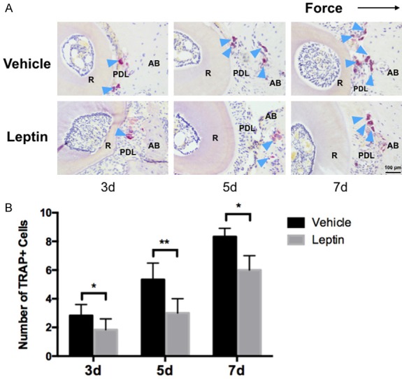
Figure 5.

Effects of leptin on osteoclatogenesis after force application in mice. A. Representative images of TRAP+ cells on the compression side of PDL and alveolar bone. B. Leptin-treated mice had significant reduced number of TRAP+ cells on 3 day and 7 day after force application. No significant difference was noted on day 5. R: root. PDL: periodontal ligament. AB: alveolar bone. The dark arrow indicated the force direction. The blue arrow indicated osteoclasts. n=5 in each group, error bar: mean ± SD. scale bar: 400 μm; **P<0.01, *P<0.05.
