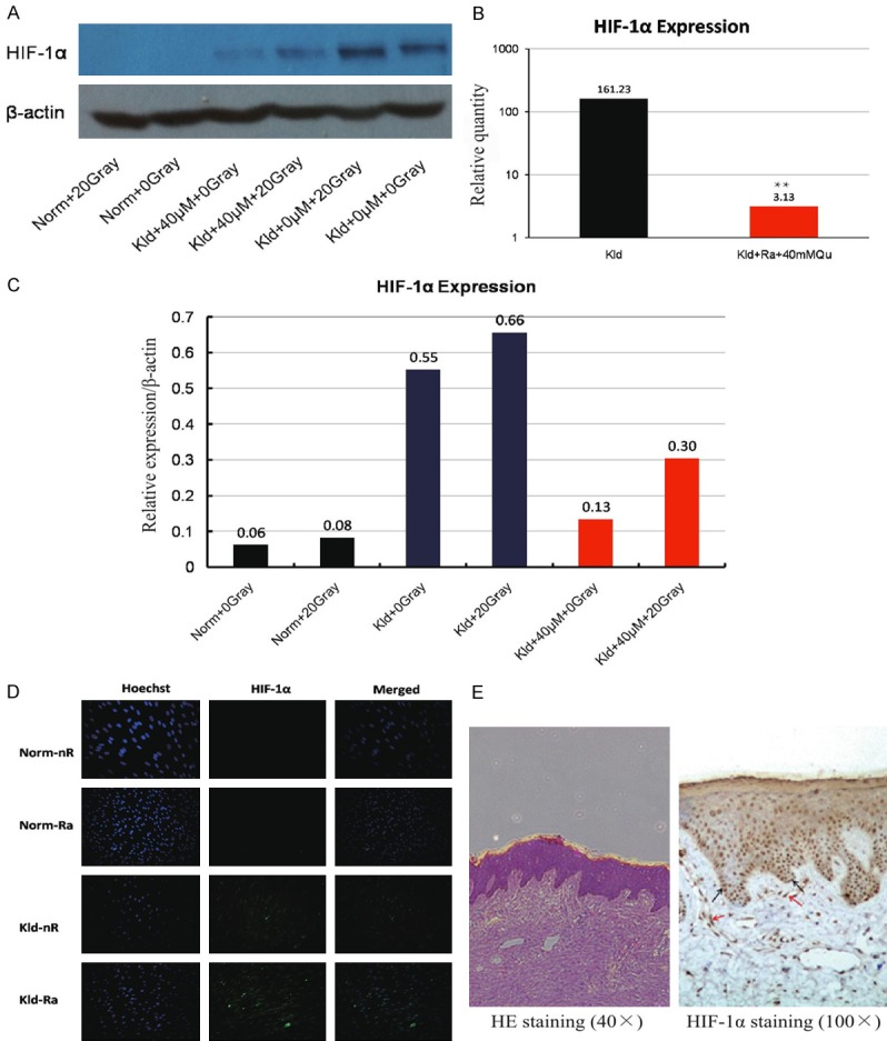
Figure 3.

HIF-1α expression in keloid fibroblasts. (A) Western blot of HIF-1α after treatment with 20 Gray IR, 40 μM quercetin, or a combination of both in normal fibroblasts and keloid fibroblasts. (B) The relative quantity of HIF-1α mRNA as measured by RT-PCR. The combination treatment of 40 μmol/L quercetin and 20 Gray IR increased the transcription of HIF-1α by 50-fold. (C) The relative expression of HIF-1α corresponding to (A). keloid fibroblasts treated with 20 Gray IR displayed a 20% increase in HIF-1α compared to non-IR treated cells, suggesting that IR may promote the expression of HIF-1α. (D) Immunofluorescence staining of HIF-1α. HIF-1α was present in the area surrounding the nucleus, and abundant in basal cells, some stratum cells, and keloid fibroblasts of the corium layer. (E) Immunohistochemistry staining of HIF-1α.
