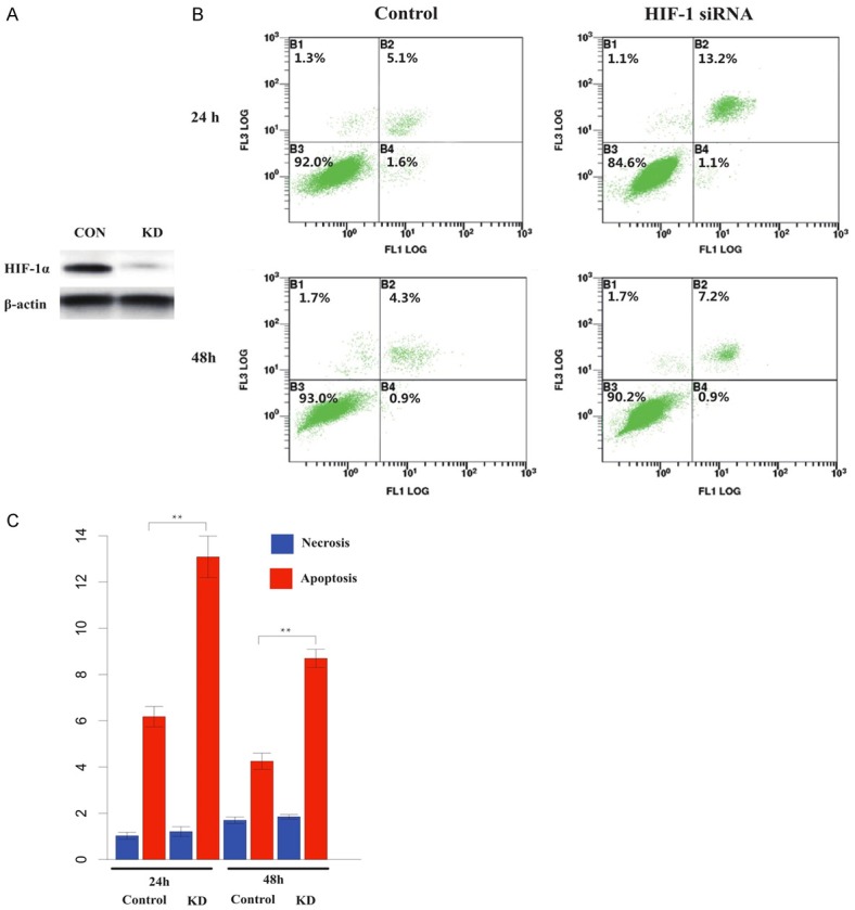
Figure 4.

Apoptosis was increased after knock-down of HIF-1α. A. Western blot of HIF-1α in keloid fibroblasts transfected with HIF-1α siRNAs (right) and control (right). B. Annexin-V flow cytometry analysis of HIF-1α deficient keloid fibroblasts and controls at 24 h and 48 h post 20 Gray IR. C. Histogram indicating percentage of necrosis and apoptosis in HIF-1α deficient keloid fibroblast. The HIF-1α deficient cells showed a substantially higher degree of apoptosis than their non-deficient counterparts at 24 h and 48 h post IR (*, P<0.05; **, P<0.01).
