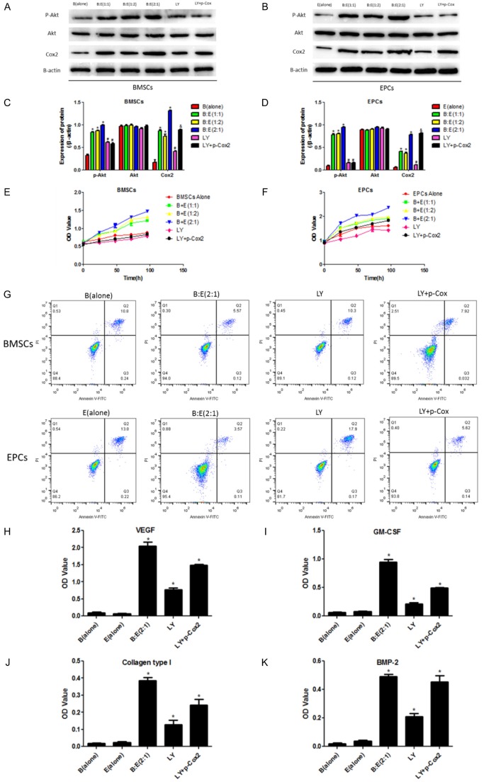
Figure 5.
Involvement of PI3K/Akt/Cox-2 axis in BMSCs and EPCs interaction mediated modulation of cell proliferation, apoptosis and angiogenesis associated cytokines secretion. A, B: Western Blot was used to detect p-Akt, Akt and Cox2 in different groups including BMSCs alone, EPCs alone, BMSCs and EPCs co-culturing system at the ratio of 1:1, 1:2 and 2:1, treating the co-culturing system (BMSCs:EPCs = 2:1) with LY294002 and synergistically treating the co-culturing system (BMSCs:EPCs = 2:1) with LY294002 and Cox-2 overexpression lentiviral vectors. The protein bands were used to quantify proteins. C, D: Image J software was performed to quantify proteins according to the grey values, the proteins were normalized by β-actin. E, F: Cell proliferation was evaluated by CCK-8 assay after treating the co-culturing system with LY294002 alone or synergistically treating the co-culturing system with LY294002 and Cox2 overexpression lentiviral vectors. The OD value was used to evaluate cell proliferative abilities. G: Flow cytometry (FCM) was employed to detect cell apoptosis rates of BMSCs and EPCs in the co-culturing system treated with LY294002 alone or synergistically treated with LY294002 and Cox2 overexpression lentiviral vectors. H-K: Detection of angiogenesis associated cytokines including VEGF, GM-CSF, Collagen type I and BMP-2 by ELISA in the culture medium of the co-culturing system treated with LY294002 alone or synergistically treated with LY294002 and Cox2 overexpression lentiviral vectors. The OD value was used to quantify the cytokines secretion. (*, P<0.05 VS B(alone) or E(alone) control groups).

