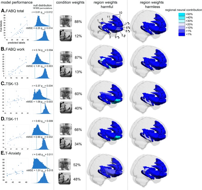
Figure 1.
The model performance (r, MSE) characterizes the strength of the relationship between true and predicted labels. Condition and region weights show the predictive contribution of the two different conditions (harmful, harmless) and fear-related brain regions (parcellated according to the AAL atlas; L, left; R, right) to the final decision function of each MKL model (questionnaires A–E with model performance; p < 0.05, FDR corrected and uncorrected). Brain regions (feature set) were identified as follows: thalamus (1); hippocampus (2); amygdala (3); insula (4); mOFC: rectus (5), Frontal_Sup_Orb (6), Frontal_Med_Orb (7); lOFC: Frontal_Mid_Orb (8), Frontal_Inf_Orb (9), mPFC: Frontal_Sup_Medial (10); anterior cingulate cortex: Cingulum_Ant (11). ← indicates the not visible contralateral homolog.

