Figure 1.
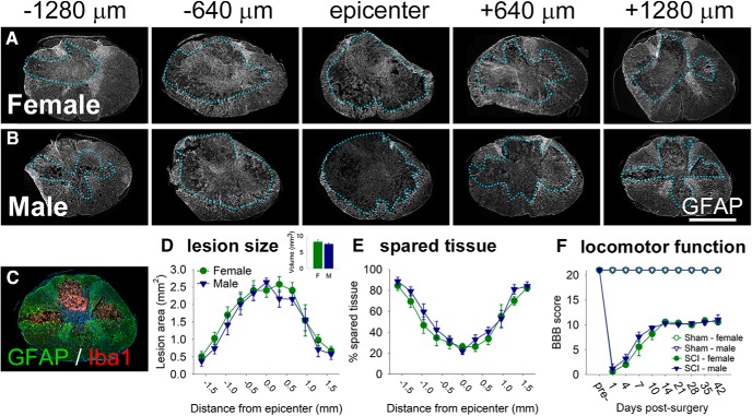
T9 contusion SCI (150 kdyn, 1 s dwell) causes extensive tissue loss at/near the epicenter with associated locomotor impairment. A, B, Spinal cords from female (A) and male (B) rats show substantial pathology and cavitation at 7 d post-SCI. GFAP immunoreactivity was used to visualize and assess tissue pathology; dotted lines outline the approximate lesion border in each section. C, Example of GFAP (astrocytes, green) and Iba1 (microglia/macrophages, red) immunoreactivity in the 7 dpi lesion site (blue, nuclei; DAPI). GFAP+ astrocytes form the glial scar; Iba1+ macrophages/microglia exist in the epicenter and Iba1+ microglia are present in the lesion penumbra. D, E, Analysis of lesion area and volume (D) and the percentage spared tissue (E) at 7 d post-SCI. There were no significant sex differences in lesion size or the percentage of spared tissue. F, Moderate T9 SCI caused substantial immediate locomotor deficits in female and male rats that recovered over time, as assessed in an open field using the BBB scale. There were no significant differences in BBB scores between female and male SCI rats. Scale bar, 1 mm.

