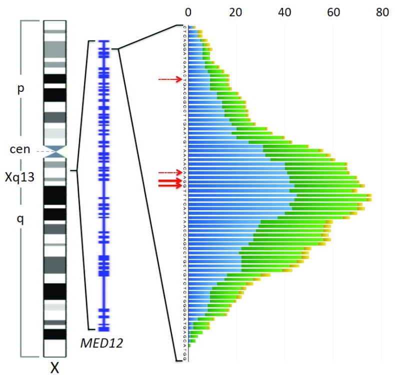
Figure 4. Patterns of MED12 deletions in uterine leiomyomas, fibroepithelial breast tumors, and chronic lymphocytic leukemia.
Left to right: Ideogram of the X-chromosome (commons.wikimedia.org), exon-intron structure of MED12 (NCBI map viewer), and plot depicting frequency of deletions at each position around the preferred site of single nucleotide exchanges (red solid arrows) seen in uterine smooth muscle tumors (blue), fibroepithelial tumors of the breast (green), and chronic lymphocytic leukemia (yellow). Deletions are plotted across all deleted base positions. Minor preferred sites of single nucleotid exchanges within exon 2 are indicated by dashed red arrows. For this diagram data on MED12 deletions from the following articles have been used: ( Guièze et al., 2015; Kämpjärvi et al., 2015; Lim et al., 2014; Markowski et al., 2013b; Mishima et al., 2015; Nagasawa et al., 2015; Ng et al., 2015; Pfarr et al., 2015; Yoshida et al., 2015) only those deletions beginning and ending in the displayed region have been considered.

