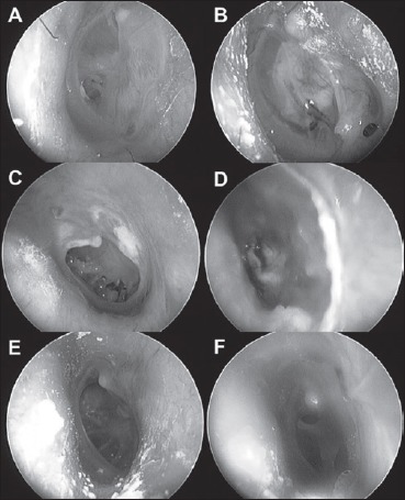
Fig. 2.

Tympanic membrane (TM) photographs in patients who underwent revision surgeries. Two patients from the native-tissue group (A-D) and one from the MegaDerm® group (F and G) underwent revision surgeries (A, C and E represent preoperative TM findings; B and F represent TM findings at 1 month postoperatively; D represents TM findings at 6 months postoperatively).
