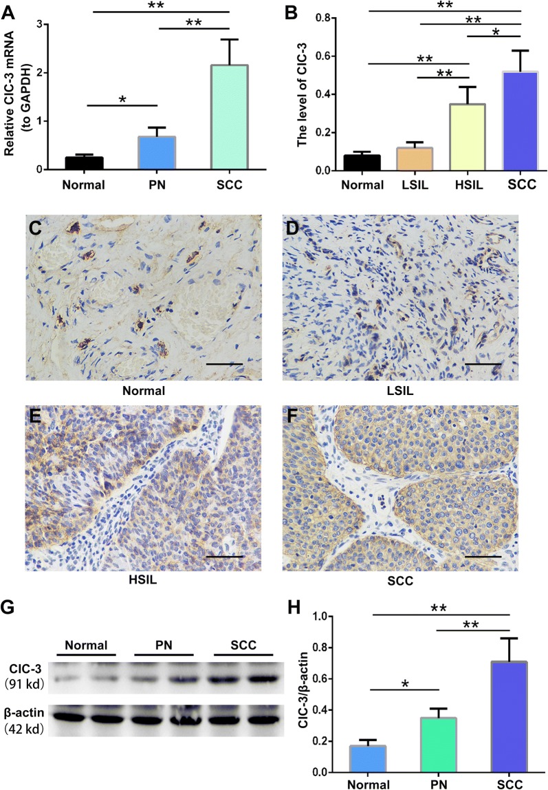
Fig. 1.
Increased expression levels of the ClC-3 mRNA and protein in paracancerous and carcinoma tissues. A ClC-3 mRNA expression levels were detected by quantitative real time RT-PCR in the control, non-cervical cancer, cervical samples (normal), the corresponding paracancerous normal tissues (PN) and the matched cancer samples (SCC) from 165 patients with cervical cancer. B Densitometric analysis of the ClC-3 protein expression levels in normal, low-squamous intraepithelial lesions (LSIL), high-squamous intraepithelial lesions (HSIL) and squamous cell carcinoma (SCC) tissues by immunohistochemistry. C–F Images of representative tissues showing the ClC-3 immunohistochemical staining. The main and inserted images were taken by a × 40 objective lens. Scale bars, 50 μm. G The representative western blot analysis of the ClC3 and β-actin proteins from normal, PN and SCC tissues. H Densitometric analysis of ClC3 protein levels in different tissues as detected by western blotting. The data in A, B and E are shown as the mean ± SD. *p < 0.05, **p < 0.01

