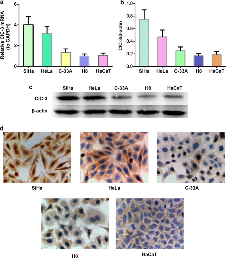
Fig. 4.
Increased expression of the ClC-3 mRNA and protein in SiHa and HeLa cell lines. a ClC-3 mRNA expression levels detected by quantitative real time RT-PCR in the SiHa, HeLa, C-33A, normal cervical epithelial cell line H8 and normal keratinocyte cell line HaCaT. b Densitometric analysis of the ClC-3 protein expression levels in SiHa, HeLa, C-33A, normal cervical epithelial cell line H8 and normal keratinocyte cell line HaCaT by western blotting. c The representative western blot of the ClC3 and β-actin proteins in the SiHa, HeLa, C-33A, H8 and HaCaT cell lines. The data in a and b are shown as the mean ± SD. d Images of the SiHa, HeLa, C-33A, normal cervical epithelial cell line H8 and normal keratinocyte cell line HaCaT, show the ClC-3 immunohistochemical staining, respectively

