Figure 1.
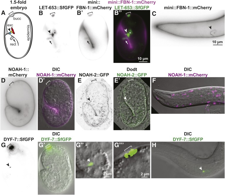
ZP proteins localize to aECMs in distinct tissues. ZP fusion proteins, expressed under their endogenous promoters, showed different patterns in elongating 1.5- to 2-fold embryos (A, B, D, E, and G) and L1 larvae (C, F, and H). All tags are inserted N-terminally to the predicted ZP cleavage site, except in the case of LET-653, where the tag is C-terminal but the cleaved product remains associated with the matrix (Gill et al. 2016). Arrowhead: excretory system; bracket: buccal cavity and nose tip; arrow: rectum. (A) Cartoon of 1.5-fold embryo, indicating major regions of aECM accumulation. Cytoplasm: white; aECM: gray; apical membranes: black. (B) LET-653::SfGFP (Gill et al. 2016) strongly lined the duct/pore lumen (Gill et al. 2016), and was also visible in or near the buccal cavity, rectum, and epidermis. Asterisk indicates neuronal coinjection marker ttx3-pro::GFP present in the FBN-1 transgene array. (B’) mini-FBN-1::mCherry (Kelley et al. 2015) strongly lined the buccal cavity, rectum, and epidermis, but was not detected in the duct/pore lumen at the 1.5- to 2-fold (n = 25) or 3-fold (n = 25) stages. (B”) Merged images illustrate distinct localization. (C) mini-FBN-1::mCherry did line the excretory duct and pore lumen in larvae. (D and D’) NOAH-1::mCherry (Vuong-Brender et al. 2017) was restricted to the region closely apposed to the epidermis and excluded from the buccal cavity, rectum, and duct/pore lumen (n = 20). (E and E’) NOAH-2::GFP (Vuong-Brender et al. 2017) was faintly visible lining the epidermis and potentially seen along the duct/pore lumen, but was never visualized at the nose tip (n = 11). F) NOAH-1::mCherry appeared intracellular in L1 larvae (n = 10). (G and G’) DYF-7::SfGFP (Low et al. 2018) localized specifically to the nose tip and a region near the duct canal junction (n = 19). (G” and G”’) show magnifications of the excretory canal (c) and nose regions, respectively. (H) DYF-7::SfGFP remained associated with the canal region in L1 larvae (n = 15).

