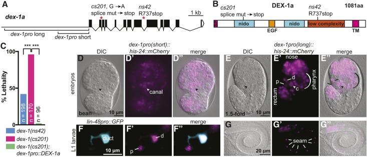
Figure 2.
dex-1 is an essential gene and broadly expressed in the embryo. (A) dex-1 promoter regions, gene structure and mutant alleles. (B) DEX-1a protein schematic and locations of mutant allele lesions. SP: signal peptide; Nido: nidogen domains; EGF: EGF-like domain. The low complexity region was previously proposed to have homology to the sperm protein zonadhesin (Heiman and Shaham 2009). We were not able to detect that similarity. TM: transmembrane domain. (C) Both ns42 and cs201 mutants show larval lethality, with a higher percentage in cs201 mutants. This lethality was efficiently rescued by a transgene encoding DEX-1a driven by the short dex-1 promoter (see A). *** P < 0.0001. Fisher’s exact test. (D and E) dex-1 transcriptional reporters were widely expressed in embryonic epithelia, including the epidermis, pharynx, nose tip, rectum, duct (d), pore (p), and canal nucleus (arrowhead, c). DIC: differential interference contrast. (D–D”) Bean stage embryo, ventral view. (E–E”) 1.5-fold stage embryo, lateral view. (F–G) dex-1 transcriptional reporter expression persisted in L1 larvae. (F–F”) Expression in the duct (labeled with lin-48pro::GFP) (d) and pore (p). (G–G”) Expression in seam epidermal cells (lines). Reporters contain either the short (D and F) or long (E and G) version of the dex-1 promoter, as shown in (A); both gave similar expression patterns.

