Figure 5.
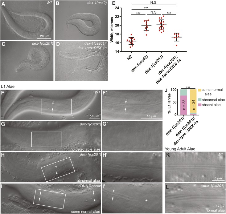
dex-1 mutants have L1 cuticle defects. (A–D) dex-1 mutants were visibly short and fat (Dpy). DIC images of newly hatched L1 larvae. (E) Quantification of hatchling width. Measurements were performed by drawing a line across the animal’s width at the region of the pharynx terminal bulb and recording line length in ImageJ. All measurements were taken three times and then averaged to find a final, recorded value. *** P < 0.0001, Mann-Whitney U-test. (F–I) DIC images of alae in newly hatched L1 larvae. (F and F’) WT L1 larvae had distinct alae, visible as two parallel ridges (arrows). Box indicates region magnified in subsequent panel. (G–H’) dex-1(cs201) L1 alae were deformed or absent. (I and I’) dex-1pro::DEX-1a transgene used to rescue lethality (see Figure 2D) rescued alae variably, with regions where alae were normal (arrows) and regions where they were missing (*). Most L1 larvae had at least some regions with normal alae. (J) Quantification of alae phenotypes. *** P < 0.0001, Freeman-Halton Fisher’s exact test. (K and L) DIC images of newly molted adults. 17/17 dex-1(cs201) mutants had normal adult alae.

