Figure 6.
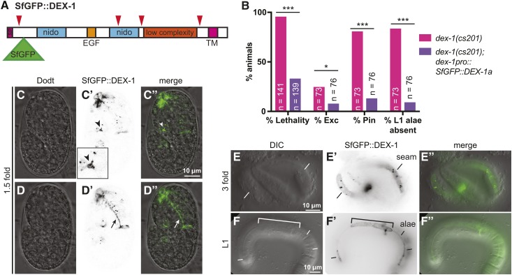
DEX-1 localizes to the aECM. (A) Model of SfGFP::DEX-1a protein showing position of SfGFP insertion after the signal peptide. Red arrowheads indicate potential cleavage sites predicted by ProP (Duckert et al. 2004). SfGFP was inserted downstream of the first predicted cleavage site in order to tag the portion of DEX-1 containing the nido and EGF domains. (B) SfGFP::DEX-1a rescued dex-1(cs201) lethality, excretory, Pin, and alae defects. *** P < 0.0001, * P < 0.01. Fisher’s exact test. (C–D”) In WT 1.5-fold embryos, SfGFP::DEX-1a localized to apical surfaces. Single Z-slices from a confocal Z-stack. (C–C”) DEX-1 was present in the duct/pore lumen (arrowhead) and particularly strong near the duct-canal junction (arrowhead). This region is enlarged in the inset. (D–D”) DEX-1 lines the pharyngeal lumen (arrow). (E–E”) threefold embryo. DEX-1 lines a strip of seam epithelium. (F–F”) In L1 larvae, DEX-1 lines alae. Bracket indicates a region of missing or abnormal alae, suggesting dominant negative effects related to inappropriately high expression level. All images shown are of WT animals expressing SfGFP::DEX-1a.

