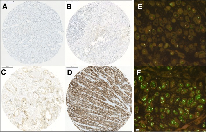
Fig. 1.
Representative images of histopathological slides to illustrate the immunohistochemistrical scoring system a-d) as well as FISH-analyses (e, f): a) Negative or staining in < 5 cells (score 0); b) very weak staining in cell groups ≥5 (score 1+); c) weak to moderate complete/basolateral/lateral staining in cell groups ≥5 (score 2+); d) strong complete/basolateral/lateral staining in cell groups ≥5 (score 3+). Representative FISH-specimens e) without and f) with HER2 amplification

