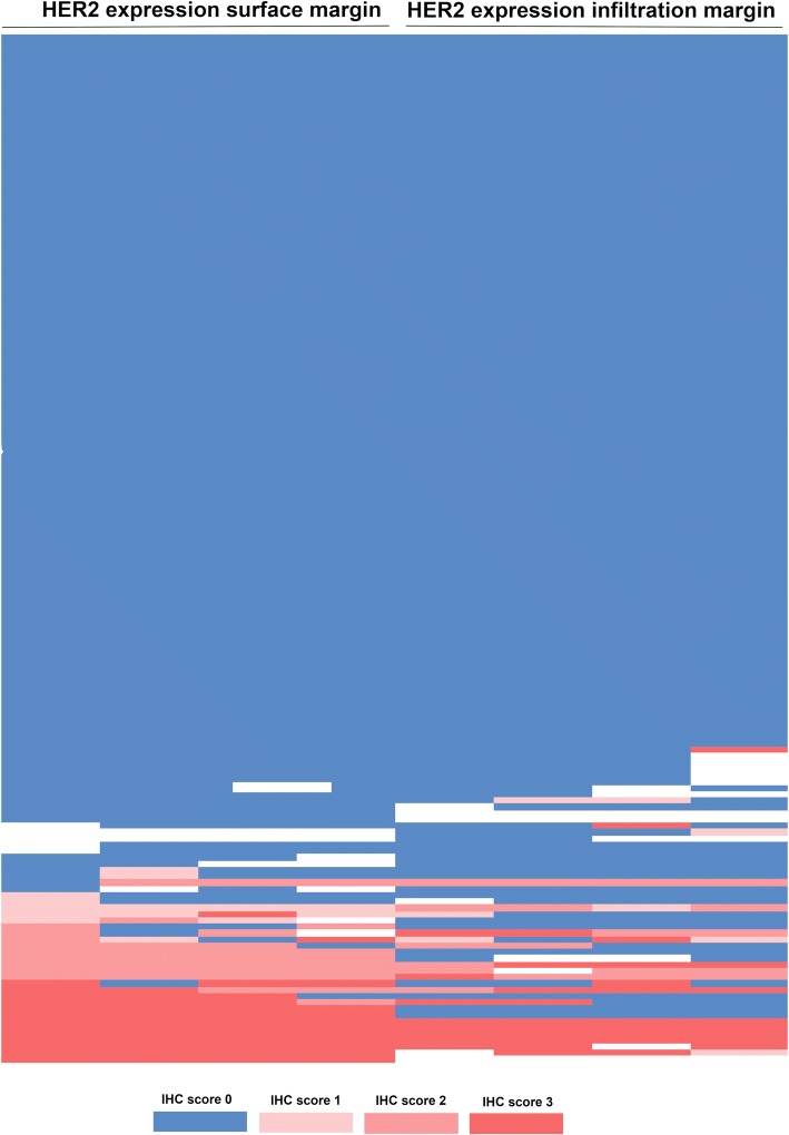
Fig. 2.
Heatmap showing heterogeneity of HER2 expression between the luminal and the infiltration area of the primary tumor. Blue area represents absence of HER2 expression (IHC score 0); light red immunohistochemistry (IHC) score 2+, FISH confirmation negative; medium red IHC score 2+, FISH positive; dark red IHC score 3 +

