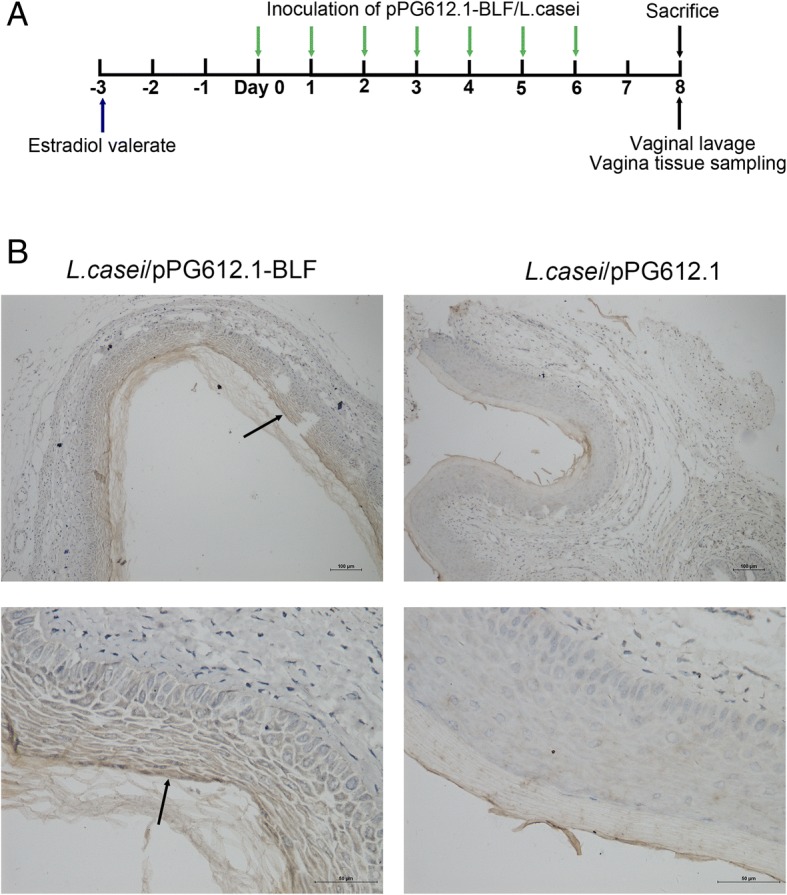
Fig. 3.

Colonization of murine vagina by L. casei/pPG612.1-BLF. a Subcutaneous injection of 100 μg of estradiol valerate was given to mice 3 days before inoculation. Twenty microliters of undiluted L. casei/pPG612.1-BLF suspension and of the two dilutions were used to inoculate the murine vaginal cavity (5 mice per group) with a pipette at the same time of day for seven consecutive days. Mice were sacrificed by cervical dislocation 2 days after the 7th inoculation; each murine vagina was washed gently with 150 μL of sterile PBS by repeated aspiration, 10 times. The vaginal tissues were fixed in 4% paraformaldehyde and embedded in paraffin prior to immunohistochemical analysis with anti-BLF primary antibodies. b A brown positive band (black arrow) was observed above the vaginal epithelial layer from mice which were inoculated with L. casei/pPG612.1-BLF into the vagina, while it was not detected in the samples from mice receiving inoculation of pPG612.1/L.casei
