Fig. 2.
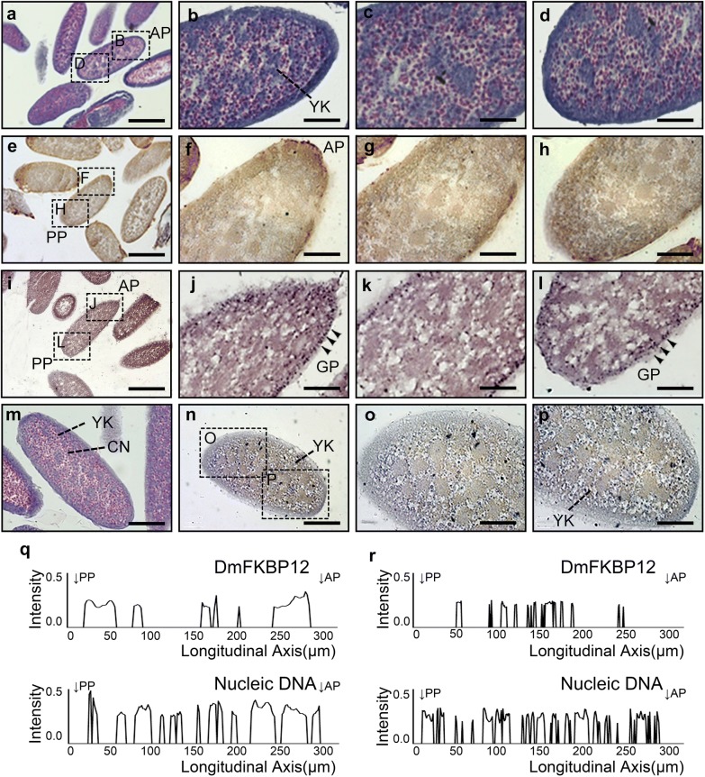
Expression profile of DmFKBP12 protein in syncytial blastoderm of the Drosophila embryo. a–d H&E staining of the syncytial blastoderm. This stage of Drosophila melanogaster embryogenesis implied 13 rapid nuclear divisions within a common cytoplasm. And these nuclear divisions produced roughly 300–400 nuclei by the end of the ninth division. e–h and n–p display the distribution of DmFKBP12 protein in the syncytial blastoderm. The DmFKBP12 was expressed in embryonic cytoplasm and plasma membrane, restricted to the periphery, anterior and posterior of the embryo before cellularization. i–l show PASM staining of syncytial blastoderm. Glycoprotein granules distribute in the periphery of the embryo. m–p presents early syncytial blastoderm in which nuclei are under division. The qualifications of DmFKBP12 distribution in early and late syncytial blastoderm are presented in q and r. Scale bar for a, e and i is 160 µm, for m, n is 80 µm, for b–d, f–h, j–l and o–p is 40 µm. AP, anterior pole of egg; PP, posterior pole of egg; YK, yolk; CN, cleavage nucleus; GP, glycoprotein

