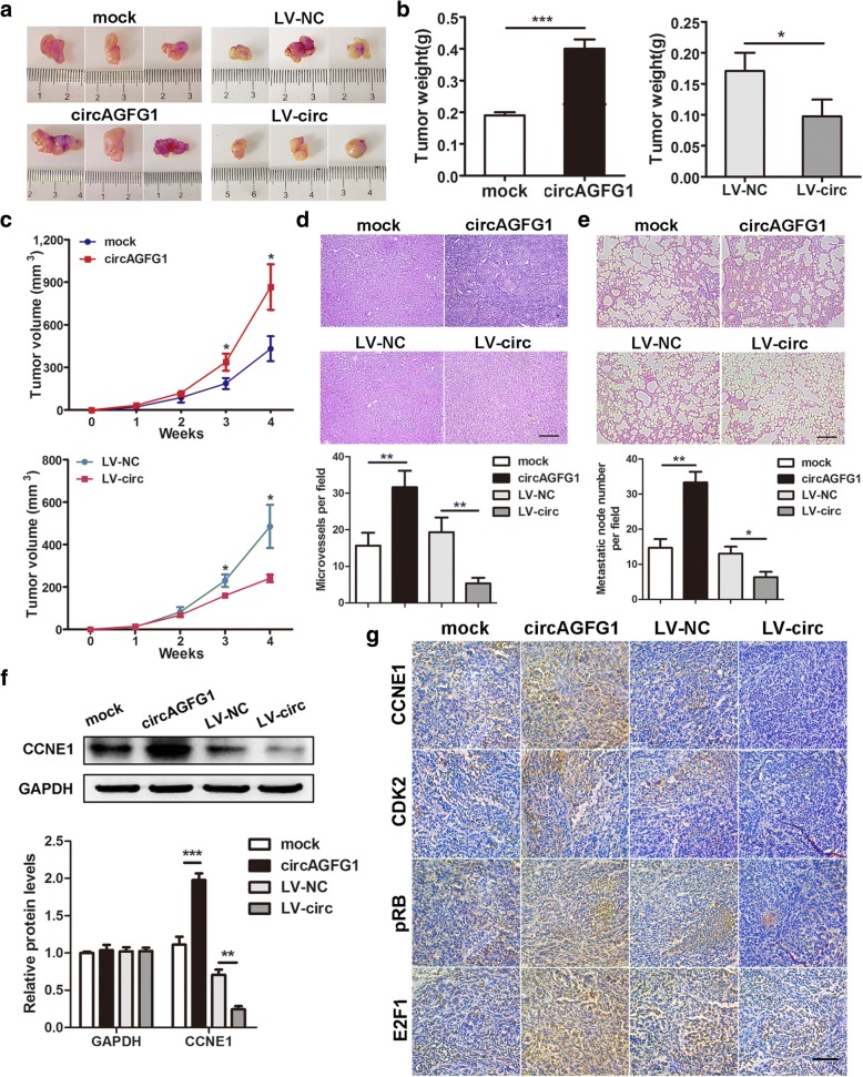
Fig. 5.
circAGFG1 facilitates tumorigenesis, angiogenesis and metastasis of TNBC cells in vivo. a Representative images of xenograft tumors of each group (n = 3). b Tumor weight was shown. c Growth curves of xenograft tumors which were measured once a week. d and e HE staining of tumor and lung sections displayed microvessels of the tumors and metastatic nodules of the lungs, respectively (magnification, × 100). Scale bar, 100 μm. f The protein level of CCNE1 was detected by western blot. g IHC staining was applied to analyze the protein levels of cell cycle-related molecules (magnification, × 200). Scale bar, 100 μm. Data were indicated as mean ± SD, *P < 0.05, **P < 0.01, ***P < 0.001

