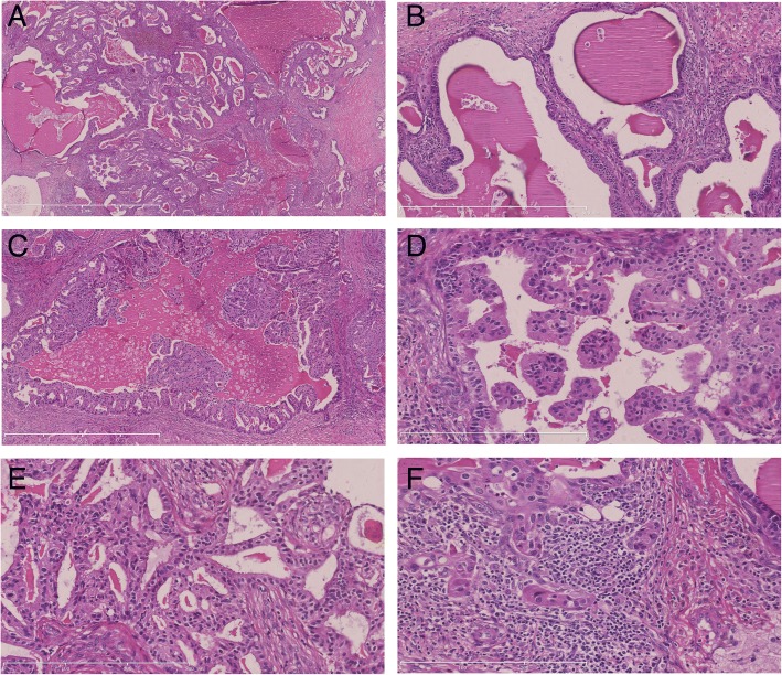
Fig. 2.
Microscopic findings. Multiple variable-sized cysts and ducts filled with thyroid colloid-like eosinophilic secretions (a, H&E, × 25). Some of the cysts are lined by flat to cuboidal epithelium (b, H&E, × 100). In other areas the epithelium showed a proliferative change in the form of pseudostratification, knobby tufts (c, H&E, × 50), micropapillary (d, H&E, × 200) and cribriform (e, H&E, × 100). An invasive component comprising of solid nests was seen (f, H&E, × 200)

