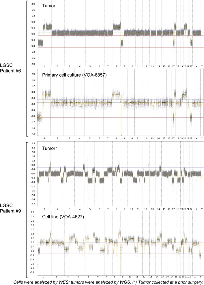
Fig. 1.
Comparison of genomic profiles between two LGSC cell cultures and their associated LGSC tumor samples. Each graph represents the copy-number (CN) changes detected per chromosome in each sample. Top graphs correspond to LGSC patient #6; CN changes detected in one of her recurrent tumor tissues was compared to the CN changes detected in the primary cell culture derived from this tissue. Bottom graphs correspond to the LGSC patient #9; CN changes detected in one of her recurrent tumor tissues was compared to the CN changes detected in the cell line established from a later recurrent tissue. High genomic profile correlation was observed between cells and tumors in both cases

