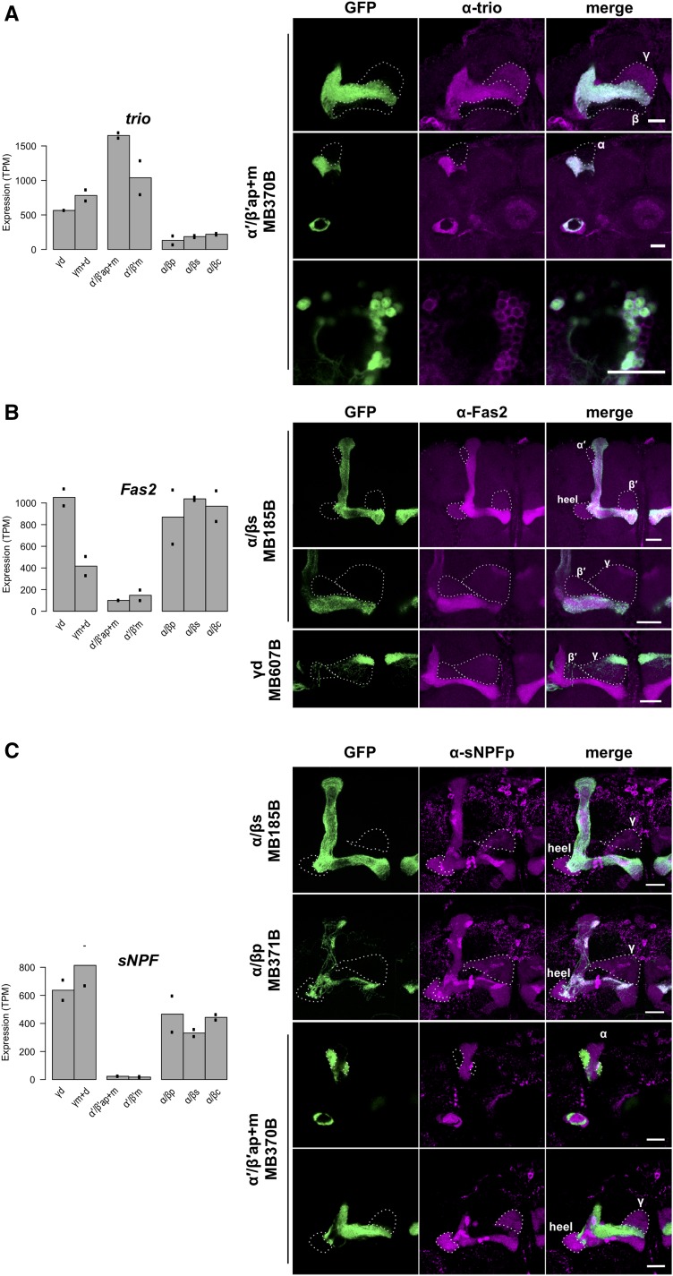
Figure 4.
(A) trio is depleted in α/β KCs. Whole mount anti-trio immunostaining confirmed strong signal in the MB α′/β′ lobes, moderate signal in the γ lobe, and no signal in the α/β lobes. The cell bodies of MB α′/β′ KC class also showed immunoreactivity. (B) Fas2 is depleted in MB α′/β′ KC class. Whole mount anti-Fas2 immunostaining confirmed stronger signal in the MB α/β and γd lobes. (C) sNPF is depleted in MB α′/β′ KC class. Whole mount anti-sNPF precursor immunostaining confirmed no detectable signal in the MB α′/β′ lobes. Among the immunoreactive α/β and γ lobes, the α/βp lobes showed the strongest signal. In each plot the bars represent the mean TPM, and the dots represent individual replicate values. Scale bars represent 20 μm. Expression patterns of the split-GAL4 lines were reported by P{10XUAS-IVS-GFP-p10}attP2.

