Figure 5.
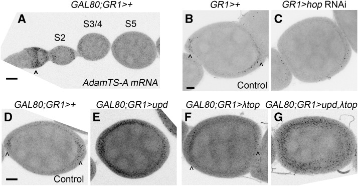
AdamTS-A expression is regulated by JAK/STAT signaling. A) Fluorescent in situ hybridization (FISH) of control (GAL4 and GAL80 only) egg chambers shows AdamTS-A expression in the germarium (arrowhead). B-G show stage 6/7 egg chambers. B) Control (GAL4 only) egg chamber for comparison with C, showing AdamTS-A expression in the anterior and posterior follicle cells (AFCs and PFCs) beginning at mid-oogenesis (arrowheads). AdamTS-A is present at reduced levels in the main body follicle cells. C) hop RNAi in the follicle cells causes a reduction in AdamTS-A expression. D) Control (GAL4 and GAL80 only) egg chamber for comparison with E-G. Arrowheads indicate AFC and PFC enrichment. E) Ectopic upd expression causes increased AdamTS-A expression in the FCs. Increased AdamTS-A expression in the main body follicle cells is particularly notable. F) Egg chambers expressing constitutively active EGFR (λtop) resemble controls. G) Egg chambers co-expressing upd and λtop show ectopic AdamTS-A expression. Panels A-G are maximum intensity projections generated from 21 z-slices at 1 μm intervals except for F, which is a projection of 20 z-slices. Scale bars represent 10 μm.

