
Fig. 1.
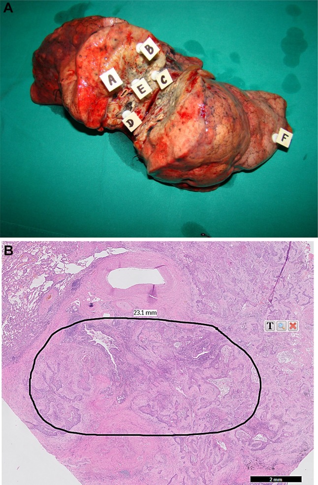
a Cross section of resected right lung with squamous cell carcinoma from a 74-year-old male patient. White squares A, B, C, D and E indicate loci of cancer tissue harvesting (samples of about 60–100 mg of wet mass). Normal lung parenchyma was collected from the place marked with square F. b Hematoxylin–eosin stained slide (fivefold magnification) prepared from a cancer tissue sample collected from locus A. The black perimeter shows the zone containing more than 90% of cancer cells which was excised for DNA isolation and KRAS mutation analysis. Slides prepared from cancer samples collected from remaining four loci revealed very similar histological images