
Figure 1:
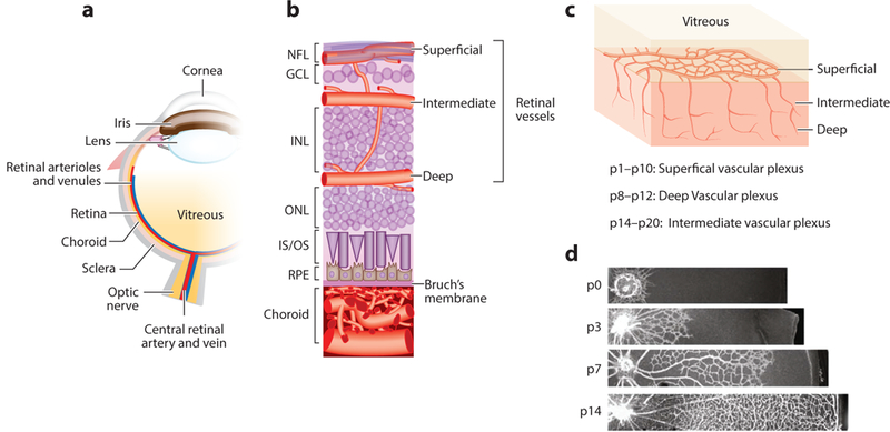
Schematic diagram of the ocular vasculature. (a) Cross-sectional image of an eye. (b) An enlarged cross-sectional view of the retinal and choroidal vasculature. Three interconnected layers of retinal vessels are embedded among inner retinal neurons: The superficial retinal vasculature lies in the NFL; the intermediate and deep retinal vascular networks lie along each side of the INL. The choroidal vessels are located beneath the RPE and Bruch’s membrane and supply oxygen and nutrients to the outer portion of the retina, which includes primarily photoreceptors in the outer nuclear layer. (c) Retinal vascular development begins with the formation of the superficial vascular plexus from pi to p10, then the deep vascular plexus from p8 to p12, and finally, the intermediate vascular plexus from p14 to p20. (d) Retinal superficial vascular plexus at different developmental stages p0, p3, p7, and p14. Panels a and b adapted from Liu et al. (2017). Panels c and d adapted from Joyal et al. (2018). Abbreviations: GCL, ganglion cell layer; INL, inner nuclear layer; IS/OS, inner segment/outer segment of photoreceptor; NFL, nerve fiber layer; ONL, outer nuclear layer; P, postnatal day; RPE, retinal pigment epithelium.