
Figure 5. Quality control of FACS sorted cell populations from the murine brain.
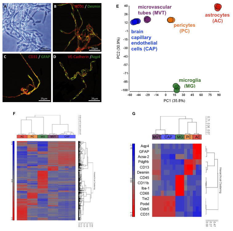
. A-D. Microvascular tubes (MVT) were isolated ( Fisher et al., 2007 ) and seeded on fibronectin-coated glass slides for double immunohistochemistry staining of brain endothelial marker CD31, pericyte marker Desmin, and astrocytic markers GFAP and Aqp4 indicating insufficient cell separation using mechanical tissue dissociation. Black scale 50 µm and white scales 25 µm. MVTs served as mixed cell population control for RNA microarray studies (E-G). E. Principal-component analysis (PCA) of the transcriptomes of brain capillary endothelial cell (CAP), pericyte (PC), astrocyte (AC), and microglia (MG) single cell populations isolated using flow cytometry. For purification of CAP and PC populations, the outlined protocol was applied, while ACs and MGs were isolated using an unpublished method. PCA was calculated for normalized EVs with a difference of at least two-fold between any pair of samples (P < 0.05 [one-way ANOVA]) and with raw expression (EV) of >140 in 100% of replicates of at least one sample population. Numbers in parentheses indicate the proportion of total variability calculated for each principal component (PC). Each data point represents a biological replicate of cells sorted from tissues pooled from 2-8 adult C57/Bl6 mice. Analysis implicates well-defined cell populations clustering together based on their cell type-specific transcriptomes. F. Hierarchical clustering by correlation of samples MVT, CAP, PC, AC, and MG with the gene list defined in E based on log2 normalized EVs. Each terminal branch represents results from a single microarray analysis of an independent biological replicate of sorted cells as described in E. G. Hierarchical clustering by correlation of selected cell-type defining gene markers: Aqp4, GFAP, and Acsa-2 (AC); Pdgfrb, CD13, and Desmin (PC); CD45, CD11b, Iba-1, and CD68 (MG); Tie2, Podxl, Cldn5, and CD31 (CAP). Individual cell types cluster together and express high levels of known cell type-specific genes indicating highly pure cell populations. As a control, arrays of MVT show the presence of pericyte, endothelial, MG and astrocyte genes.