Figure 2.

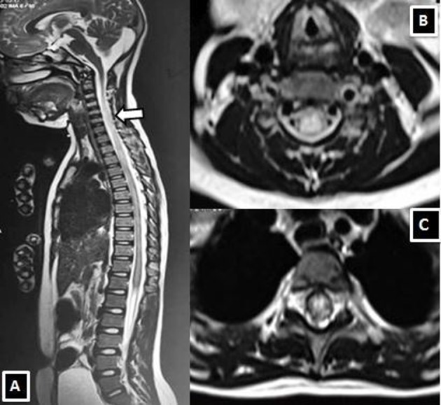
MRI spine T2-weighted sagittal (A) and T2 weighted axial images at mid-cervical (B) and thoracic spinal cord (C) demonstrating longitudinally extensive transverse myelitis in the cervicodorsal cord (white arrow).

MRI spine T2-weighted sagittal (A) and T2 weighted axial images at mid-cervical (B) and thoracic spinal cord (C) demonstrating longitudinally extensive transverse myelitis in the cervicodorsal cord (white arrow).