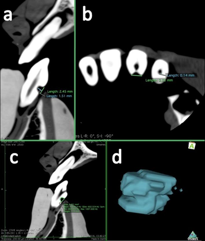
Figure 4.
Representative OsiriX analysis software images from a scan obtained at 0.2 mm voxel size of a mandibular anterior tooth with artificial cervical ERR cavity. (a) Shows a sagittal section providing the height and depth of the cavity, (b) shows an axial section providing the diameter of the cavity and (c) shows sagittal sections illustrating the segmentation process for volume quantification. Observers generated volume renderings by creating the region of interest with a pencil tool. ERR, external root resorption.

