
Figure 6.
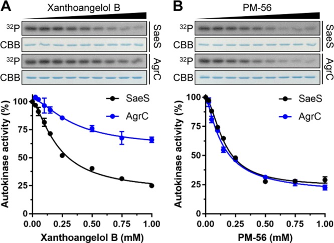
Inhibitory effects of xanthoangelol B 1 and PM-56 on the in vitro autokinase activities of SaeS and AgrC. Purified recombinant minimal kinase domains of SaeS and AgrC (5 μM each) were incubated at various concentrations (0–1 mM) of 1 (A) or PM-56 (B) for 15 min on ice before addition of ATP to initiate the autophosphorylation reactions. After SDS-PAGE analysis, total and 32P-labeled proteins were visualized by Coomassie-Brilliant Blue staining and autoradiography (32P), respectively. Each lane contains approximately 0.5 μg purified SaeS or AgrC. The 32P-labeled protein bands were quantified by densitometric analysis, and the autokinase activities were calculated as the percentages of 32P-labeled proteins in the samples treated with inhibitory chemicals compared to those in the control samples treated with 10% DMSO. Dose–response curves were generated by plotting the percent autokinase activity against the chemical concentration. The experiments were conducted in duplicate.