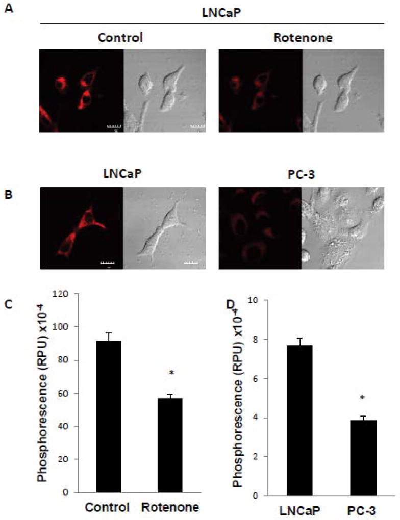
Figure 1. Mitochondrial respiratory function regulates mitochondrial oxygen concentration.
(A) LNCaP cells were incubated in the absence or presence of 2 µM rotenone for 6 h and mitochondrial oxygen level monitored by mitoBTP, as described in Materials and Methods. Representative cells are shown with the red signal indicating mitoBTP phosphorescence and differential interference contrast microscopy showing corresponding whole cell morphology. The scale bar indicates 10 µm. (B) Measurement of mitochondrial oxygen level by mitoBTP in LNCaP and PC-3 cells. Other details as in panel A. (C, D) Quantitation of mitoBTP phosphorescence from panel A and panel B, respectively. Data were derived from the examination of 10 cells/condition and are shown as mean ± S.D. * p<0.001.

