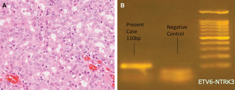
Fig. 2.

A, Histopathological image of the surgical specimens (HE staining ×100). From the cyst wall, tumor cells grow within the vascular fibrotic stroma forming a microcystic structure. B, Genetic analysis by PCR revealed ETV6-NTRK3 gene fusion. PCR, polymerase chain reaction.
