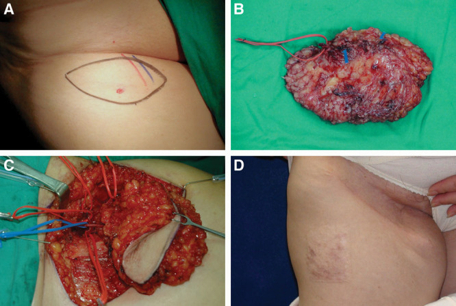
Fig. 1.

A 65-year-old woman who was a victim of right breast cancer postmastectomy, axillary lymph node dissection, and chemoradiation. She suffered from right upper limb lymphedema with 3 episodes of cellulitis per year for 2 years. She underwent right vascularized groin lymph node flap transfer to right dorsal wrist. Skin paddle 12 × 6 cm was designed on right groin below the inguinal ligament and close to common femoral vessels. One perforator was marked with pencil of medial Doppler (A). The superficial circumflex vessels were identified with vessel loop. The flap was elevated with short pedicle artery and 2 veins (C). The donor site of right groin 6 years after (D).
